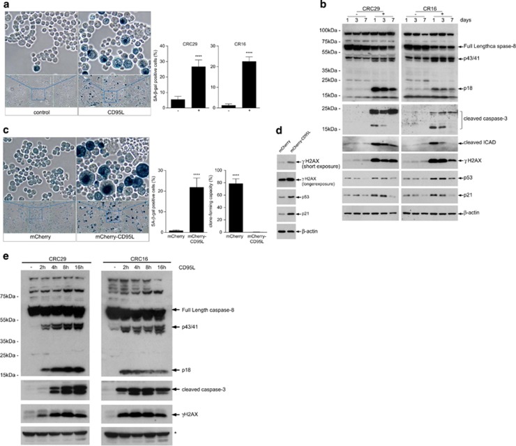
Figure 3.
CD95L induces senescence in human colonospheres. (a). Human colonospheres (CRC29, CR16) were either treated with FC control or with 10 ng/ml of FC-CD95L for 7 days. Control and CD95L-treated cells were then analyzed for SA-βGAL activity. Significance (unpaired t-test): P<0.0001; CR16 P<0.0001. (b) Human colonospheres (CRC29, CR16) were treated with FC control (−) or 5ng/ml FC-CD95L (+) for 1, 3, or 7 days. Western blot analysis of caspase-8 and -3 activation, cleavage of iCAD, and the DNA damage marker γH2AX over time. (c) A lentiviral vector for expression of mCherry-CD95L or a control vector (mCherry backbone without CD95L) was introduced into human CRC29 colonospheres. Ten days after transduction, SA-βGAL activity was measured as in a. Significance (unpaired t-test): βGAL assay P<0.0001; colony-forming assay: P<0.0001. (d) CRC29 cells expressing either mCherry or mCherry-CD95L (as in c) were analyzed by western blotting for the presence of γH2AX, p53, and p21 induction. (e) Human colonospheres (CRC29, CR16) were either treated with FC control (−) or were with 10 ng/ml of FC-CD95L for the indicated time points. Western blot analysis was then used to assess caspase-8 and -3 activation and the DNA damage marker γH2AX over time. * indicates unspecific band

