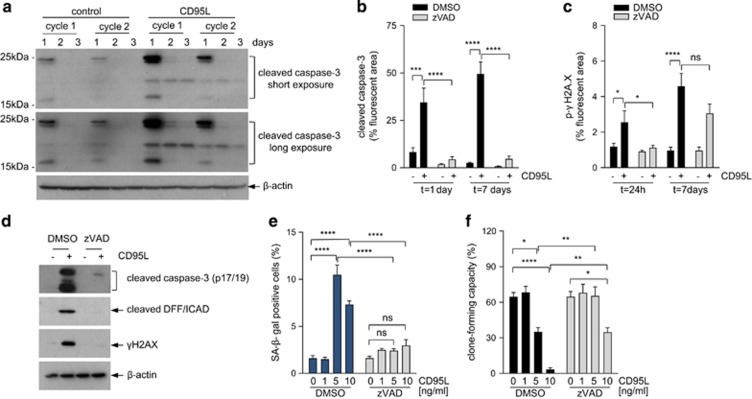
Figure 4.
CD95-induced senescence is mediated by low-level canonical caspase signaling. (a) CRC29 colonospheres were exposed once (cycle 1) or chronically exposed to CD95L (10 ng/ml) for 2 weeks (cycle 2), and collected at the indicated time points. Cells were collected and analyzed for the presence of activated (cleaved) caspase-3 by western blotting. (b–c) CRC29 colonospheres were exposed to CD95L (5 ng/ml) in the presence or absence of zVAD (25 μM) for 1 or 7 days and were then processed for imunofluorescence analysis of caspase-3 activation (b) and DNA damage (γ-H2AX) (c) An overview of the significance of all comparisons is provided in Supplementary Table S1. (d) Colonspheres were exposed to CD95L (10 ng/ml) for 24 h in the presence or absence of zVAD (25 μM) followed by western blot analysis of caspase cleavage and iCAD processing. (e and f) CRC29 colonospheres were exposed to increasing concentrations of CD95L as indicated in the presence or absence of zVAD (25 μM), after which SA-βGAL (e) and clone-forming capacity (f) were assessed at 7 or 14 days, respectively. The asterisks indicate significant differences (ordinary one-way ANOVA) (****P<0.00001; **P<0.001). An overview of the significance of all comparisons is provided in Supplementary Table S1

