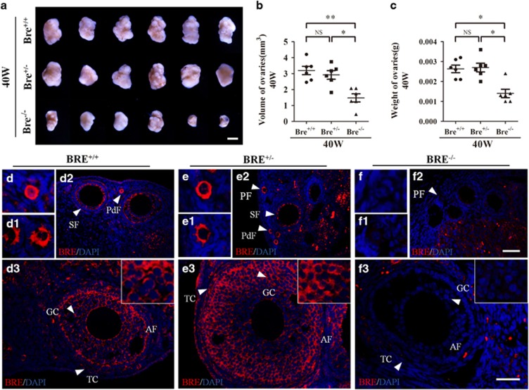
Figure 1.
BRE knockout mice produce smaller ovaries. (a) Representative appearance of 40-week-old BRE+/+, BRE+/− and BRE−/− ovaries. (b and c) Plot charts comparing the ovarian volumes (b) and weights amongst the three groups of ovaries. (d–f) Representative immunofluorescent micrographs of BRE+/+ (d), BRE+/− (e) and BRE−/− (f) ovarian sections stained with BRE antibodies. The staining confirms that BRE−/− mutant primordial (f), primary (f1), secondary (f2) and antral (f3) follicles do not express BRE. Furthermore, in BRE+/+ and BRE+/− follicles, BRE is manly expressed in the granulosa cells (d and e). PdF, primordial follicle; PF, primary follicle; SF, secondary follicle; TC, theca cells; GC, granulosa cells. Scale bars=1000 μm in a; 50 μm in d2, e2 and f2; 50 μm in d3, e3, f3

