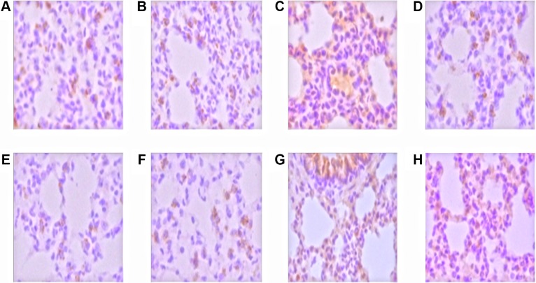
Figure 2. The expression of p38MAPK and ERK in the nucleus was stained with brown and yellow.
Control group (A) and GLA group (B), the p38MAPK expression was weak and scattered in the airway epithelium and alveolar epithelial cells; LPS group (C), p38MAPK expressed in inflammatory cells, alveolar epithelial cells, airway epithelial cells and vascular endothelial cells; LPS+GLA group (D), p38MAPK expressed cells decreased significantly. Control group (E) and GLA group (F), ERK expression was weak, distributed in the cytoplasm of airway epithelial cells and alveolar epithelial cells; LPS group (G), ERK expressed in inflammatory cells, alveolar epithelial cells, airway epithelial cells and vascular endothelial cells; LPS+GLA group (H), ERK expressed cells decreased significantly. (original magnification ×400)

