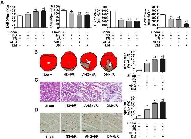
Figure 1. Hyperglycemia exacerbated cardiac dysfunction, increased infarct size and apoptosis index after myocardial ischemia/reperfusion in rats.

A. LVEDP, LVESP, +LVdp/dtmax and -LVdp/dtmax. LVEDP, left ventricular end diastolic pressure; LVESP, left ventricular end systolic pressure; +LVdp/dtmax, left ventricular maximal rate of pressure increase; -LVdp/dtmax, left ventricular maximal rate of pressure decrease. B. Left: representative photographs of heart sections stained by 2, 3, 5-triphenyltetrazolium (TTC); Right: myocardial infarct size expressed as percentage of the total left ventricular area. C. Representative photographs of myocardial tissue with H&E staining. Scale bar, 200μm. D. Left: representative photographs of in situ detection of apoptotic myocytes by TUNEL staining; Right: percentage of TUNEL-positive nuclei in heart tissue sections. Scale bar, 50μm. Sham, sham-operated; NS, normal saline; I/R, myocardial ischemia/reperfusion; AHG, acute hyperglycemia; DM, diabetes. Data are means ± SEM of 8 to 10 samples in each group. *P<0.05 vs Sham group; †P<0.05 vs NS+I/R group.
