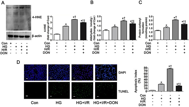
Figure 6. O-GlcNAc modification of ALDH2 contributed to 4-HNE accumulation, aldehydes and carbonyl formation and apoptosis.

A. The level of 4-HNE-protein adducts in Con, HG, HG+H/R, HG+H/R+DON groups in vitro. Cell lysates were separated by SDS-PAGE, and western blot was performed using specific anti-4-HNE antibody. B. The level of aldehydes. C. The level of protein carbonyl formation. D. Left: representative photographs of in situ detection of apoptotic cells by TUNEL staining; Right: percentage of TUNEL-positive nuclei in H9c2 cells. Scale bar, 200μm. Mannitol was used to keep the osmolarity consistent among different glucose concentrations in vitro. Con: normal glucose; HG: high glucose; H/R: hypoxia/reoxygenation; DON: a specific O-GlcNAc modification inhibitor. Data are means ± SEM of 4 to 5 samples in each group. *P<0.05 vs Con group, †P<0.05 vs HG group, ‡P<0.05 vs HG+H/R group.
