
Figure 7. NF exosomes transfected with miRs -21, -143, -378e, similarly to CAF exosomes, promote stemness and epithelial-mesenchymal transition phenotype.
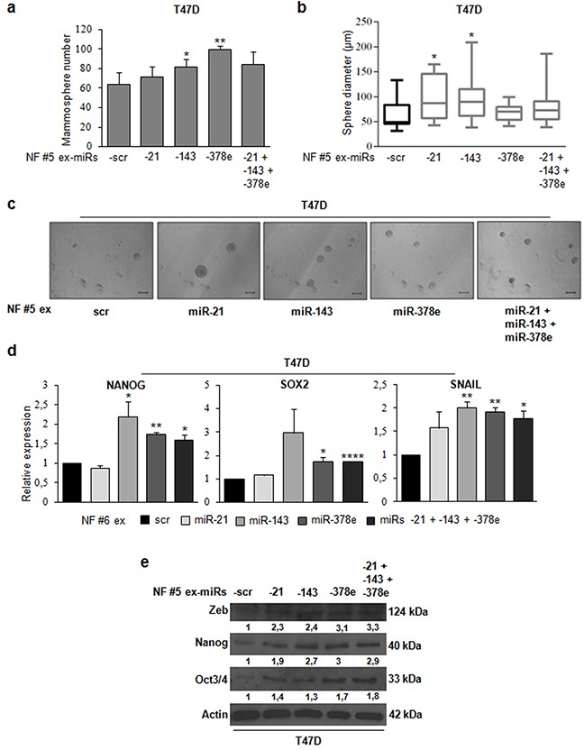
NF (patients #5, and #6) were cultured for 48h. Then, exosomes were isolated from NF-conditioned media with ExoQuick-TC™ solution, and transfected using Exo-Fect™ solution with scrambled miRs (scr, control), or miRs -21, -143, -378e, alone or in combination (final concentration: 130nM). T47D cells were cultured in the presence of NF exosomes (patient #5) transfected with either scrambled miRs (control) or miRs for 48h. Cells were harvested and grown under non-adherent conditions in stem medium. After four days, the capacity of cells to form spheres was assessed a. Sphere diameter distribution for 10 representative fields b. Scale bar: 100μm c. In addition, T47D cells were cultured in the presence of NF exosomes (patients #5, and #6) transfected with either scrambled miRs (control) or miRs for 24h d or 48h e. Real Time PCR was performed to analyze nanog, sox2, and snail mRNA (d). Western blot analysis was performed to evaluate zeb, nanog, and oct3/4 protein levels. Western blot analysis from representative experiments. Actin was used as loading control. The experiments were repeated at least three times (e). In a, b, d, data were obtained from three independent experiments and are presented as mean value ± SD. P-value calculated using Student's t test. * p<0.05; ** p<0.01; *** p<0.001; **** p<0.0001 (over control).