
Figure 5. Effects of DAC on PTEN/AKT pathway in TE13 and YES-2 cells.
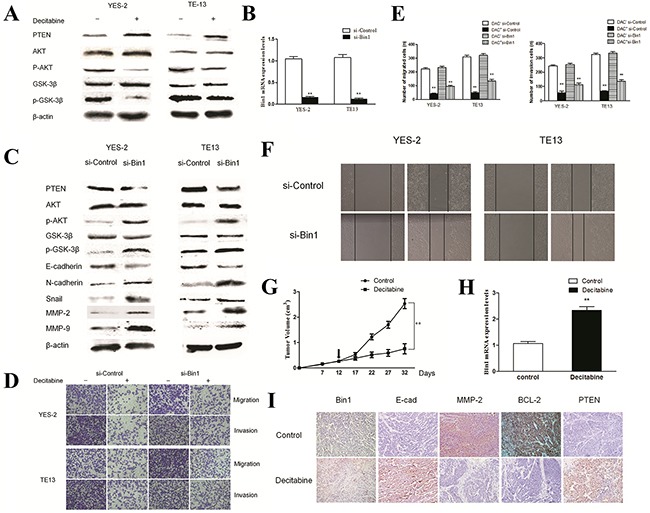
A. Effect of DAC on the PTEN/AKT pathway. YES-2 and TE13 cells were treated with DAC at concentrations of 90 μM. The expression of PTEN, AKT and GSK-3β proteins were detected by western blot. β-actin was used as an internal control. The figures are representative examples of three independent experiments. B. Effect of Bin1-siRNA on the expression of Bin1 mRNA expression in DAC-treated YES-2 and TE13 cells by qRT-PCR. C. Effect of Bin1-siRNA on the expression of PTEN and AKT proteins in DAC-treated YES-2 and TE13 cells. DAC-treated (90 μM) YES-2 and TE13 cells were transfected with Bin1-siRNA. The related proteins were detected by western blot. β-actin was used as an internal control. The figures are representative examples of three independent experiments. D. and E. Effect of Bin1-siRNA on the migration and invasion of DAC-treated YES-2 and TE13 cells. DAC-treated (90 μM) YES-2 and TE13 cells were transfected with Bin1-siRNA. After 48 h, the migration and invasion were observed using a transwell migration assay. The number of invading cells was calculated (n = 3). F. Effect of Bin1-siRNA on wound healing of DAC-treated YES-2 and TE13 cells for 48 h. DAC-treated (90 μM) YES-2 and TE13 cells were transfected with Bin1-siRNA. G. The tumor volume of nude mice with or without DAC. H. The expression level of Bin1 mRNA in nude mice with or without DAC. I. Representative immunohistochemistry images showing expression statuses of Bin1, E-cadherin, MMP2, BCL-2 and PTEN in tumor tissues of nude mice received different treatment (×200). ** P < 0.01.