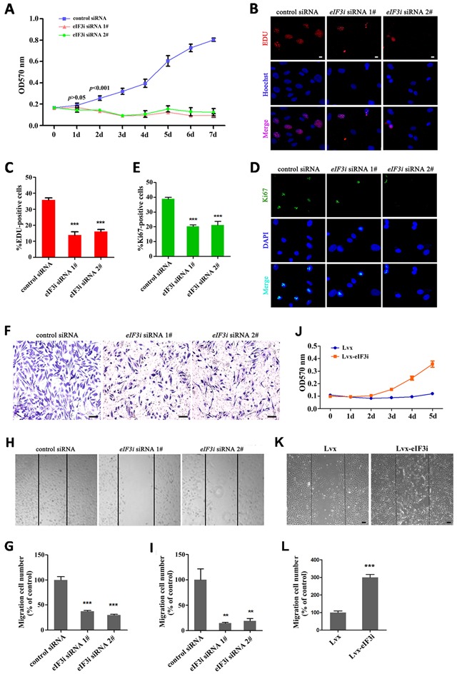
Figure 3. eIF3i promotes vascular endothelial cells proliferation and migration.

A. eIF3i siRNAs suppressed the HUVEC viability. MTT, 3-(4,5-dimethyl-2-thiazolyl)- 2,5-diphenyl-2-H-tetrazolium bromide (n=2). B. EDU assays in control siRNA- and eIF3i siRNAs-transfected HUVECs. Scale bars, 10μm. C. The statistics of EDU positive cells. (n=3). ***p< 0.001. D. Ki-67 immunofluorescent staining in control siRNA- and eIF3i siRNAs-transfected HUVECs. Scale bars, 10μm. E. The statistics of Ki-67 positive cells. (n=3). ***p<0.001. F. Transwell assay of control siRNA- and eIF3i siRNAs-transfected HUVECs. Scale bars, 100μm. G. The statistics of invaded cells in transwell assay. H. eIF3i siRNAs inhibited HUVECs migration in heal wound assay. Scale bars, 100μm. I. The statistics of cell migration. (n=3). **p<0.01. J. eIF3i overexpression increased the HUVEC viability. (n=3). K. eIF3i overexpression increased HUVECs migration in heal wound assay. Scale bars, 100μm. L. The statistics of cell migration. (n=3). ***p< 0.001.
