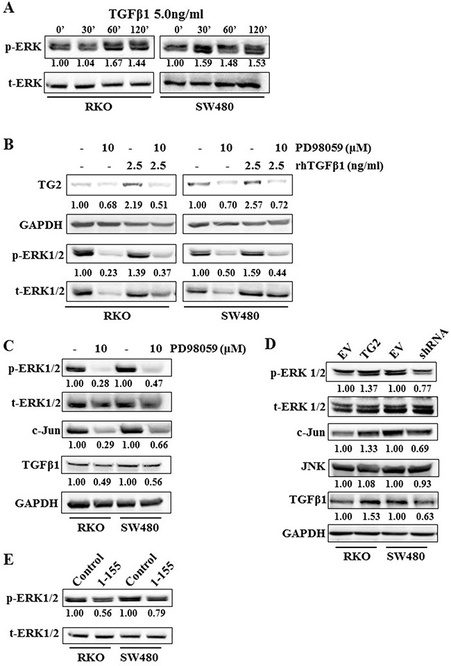
Figure 5. ERK1/2 plays a role in TGFβ1 induced TG2, and TG2 induced TGFβ1 expression in RKO and SW480 cells.

A. Western blotting showing ERK1/2 activation, after TGFβ1 (5.0 ng/ml) treatment over a time course of 2 h in wt RKO and SW480 cells. B. Western bloting of whole cell lysates of wt RKO and SW480 cells, showing expression of TG2 and ERK1/2 after treatment with TGFβ1 with or without ERK inhibitor PD98059 (10 μM). C. Western blotting of whole cell lysates from wt RKO and SW480 cells showing expression of ERK1/2, C-Jun and TGFβ1 with or without treatment of cells with ERK inhibitor PD98059 (10 μM). D. Western blotting of whole cell lysates of RKO and SW480 control cells and cells transduced with TG2 (RKOTG) or shRNA (SW480shRNA), showing TG2 expression, ERK1/2 activation and TGFβ1 expression. E. Western blotting of whole cell lysates showing activation of ERK1/2 in wt RKO and SW480 cells with or without treatment with TG2-sepcific cell permeable inhibitor 1-155 inhibitor (1 μM).
