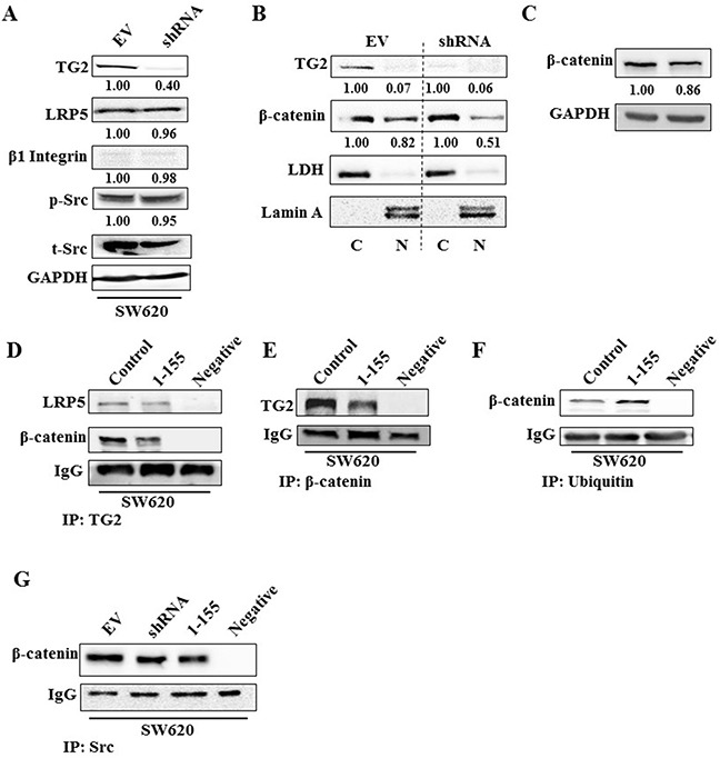
Figure 6. TG2 interacts with LRP5 prevents ubiquitination of β-catenin and induces β-catenin accumulation in the nucleus.

A. Western blotting of whole cell lysates of SW620 EV control cells and SW620 transduced with TG2shRNA (SW620shRNA), showing expression of TG2, LRP5, β1 Integrin, phosphorylated and total Src. B. Western blotting of β-catenin presence in the nuclear (N) and cytoplasmic (C) in cell extracts of SW620 control cells and SW620shRNA cells. LDH and Lamin A were used as cytoplasmic and nuclear protein markers respectively. The N and C fractions were separated as described in the Materials and Methods. C. Western blotting of β-catenin expression in whole cell lysates of SW620 control cells and SW620shRNA cells. D. Western blotting of LRP5 and β-catenin in wt SW620 treated with DMSO (control) and SW620 cells treated with TG2 selective inhibitor 1-155 (1 μM), following TG2 co-IP from whole cell lysates. PBS containing no cell lysates was used as the negative control sample. Co-IP was undertaken as described in the Materials and Methods. E. Western blotting of whole cell lysates for TG2 after β-catenin co-IP, following treatment of cells with TG2-selective inhibitor 1-155 (1 μM) or DMSO control. PBS containing no cell lysates was used as the negative control sample. F. Western blotting of whole cell lysates from wt SW620 cells after treatment with TG2 selective inhibitor 1-155 (1 μM) or DMSO control, showing β-catenin after ubiquitin co-IP of whole cell lysates from wt SW620. PBS containing no cell lysates was used as the negative control sample. G. Western blotting of β-catenin after Src co-IP from whole cell lysates of SW620 EV cells (treated with DMSO) and SW620 shRNA cells or after treatment with TG2 inhibitor 1-155. PBS containing no cell lysates was used as the negative control sample.
