
Figure 3. Biphasic activation of A-SMase in CD95-receptosomes.
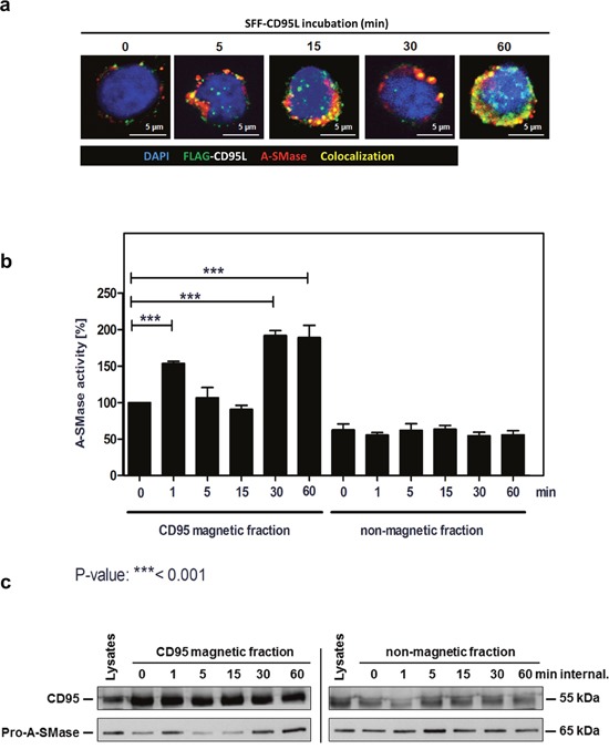
a. Merged confocal microscopic images of SKW6.4 cells stimulated with 100 ng/ml SFF-CD95L. The CD95L was visualized by an anti-FLAG antibody and anti-mouse Alexa-Fluor 488. A-SMase stained by a polyclonal anti-A-SMase antibody and anti-rabbit Alexa-Fluor 555 was detected in CD95-receptosomes between 1 min and 60 min internalization. Co-localization of CD95 and A-SMase is indicated in yellow. b. Magnetic and non-magnetic fractions were used to measure A-SMase activation in SKW6.4 cells after indicated time points of CD95 internalization. Three independent preparations were pooled and measured in triplicate. (1way-Anova with Bonferroni's multiple comparison test) c. Western blot analysis of CD95 magnetic and non-magnetic fractions with anti-CD95 and anti-A-SMase antibodies. Corresponding to the activity assay, Western blot analysis demonstrated a biphasic appearance of A-SMase in the magnetic fractions.