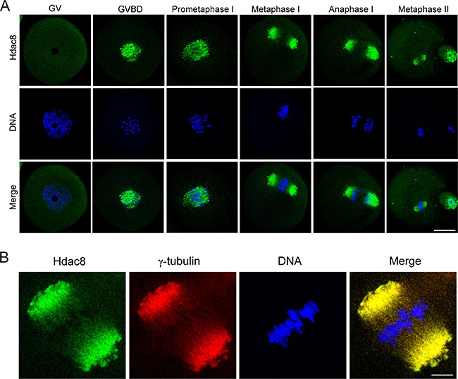
Figure 1. Subcellular localization of HDAC8 during mouse meiotic maturation.

A. Mouse oocytes at GV, prometaphase I, metaphase I, anaphase I and metaphase II stages were immunolabeled with anti- HDAC8 antibody (green) and counterstained with Hoechst (blue). Images were acquired under the confocal microscope. Scale bar, 20 μm. B. Metaphase I oocytes were double-stained with anti-HDAC8 antibody (green) and anti-γ-tubulin antibody (red) and then counterstained with Hoechst (blue). Scale bar, 5 μm.
