
Figure 6. Depletion of HDAC8 disrupts the localization of γ-tubulin in mouse oocytes.
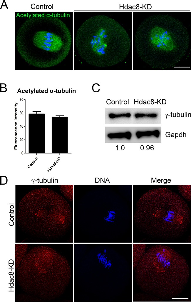
A. Representative images of acetylated α-tubulin in control and HDAC8-KD oocytes. Oocytes were immunostained with anti-acetylated α-tubulin antibody and counterstained with Hoechst to visualize chromosomes. Scale bar, 20 μm. B. The immunofluorescence intensity was recorded in control and HDAC8-KD oocytes. Data were presented as mean percentage (mean ± SEM) of at least three independent experiments. Asterisk denotes statistical difference at a p < 0.05 level of significance. C. Protein levels of γ-tubulin in control and HDAC8-KD oocytes. The blots were probed with anti-γ-tubulin antibody and anti-Gapdh antibody, respectively. D. Localization of γ-tubulin in control and HDAC8-KD oocytes. Oocytes were immunostained with anti-γ-tubulin antibody and counterstained with Hoechst. Scale bar, 20 μm.