
Figure 3.
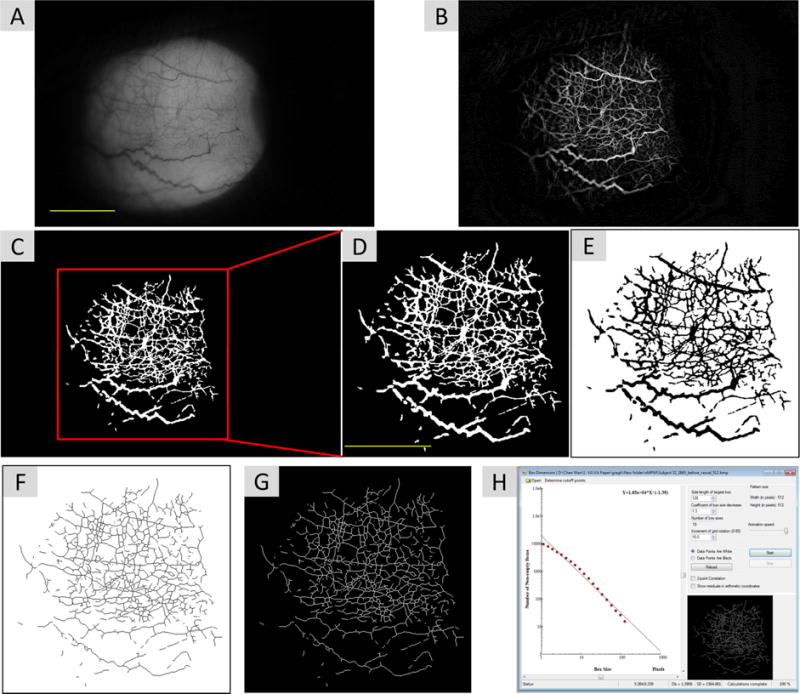
Non-invasive microvascular perfusion maps (nMPMs) and fractal analysis. Custom software was developed to segment conjunctival vessels and to create the nMPMs for fractal analysis using a series of image processing procedures. The raw image was first resized from 3,456 × 2,304 pixels to 1,024 × 683 pixels (A). Imaging processing using morphological opening was performed to create nMPMs (B). Vessels were segmented (C) and the image was cropped a field of view 7.85 × 7.85 mm2 containing 512 × 512 pixels (D). The cropped image was then inverted (E) and skeletonized (F). The image was further inverted back for fractal analysis (G) using monofractal analysis (i.e. box-counting, H). Bars=3 mm.