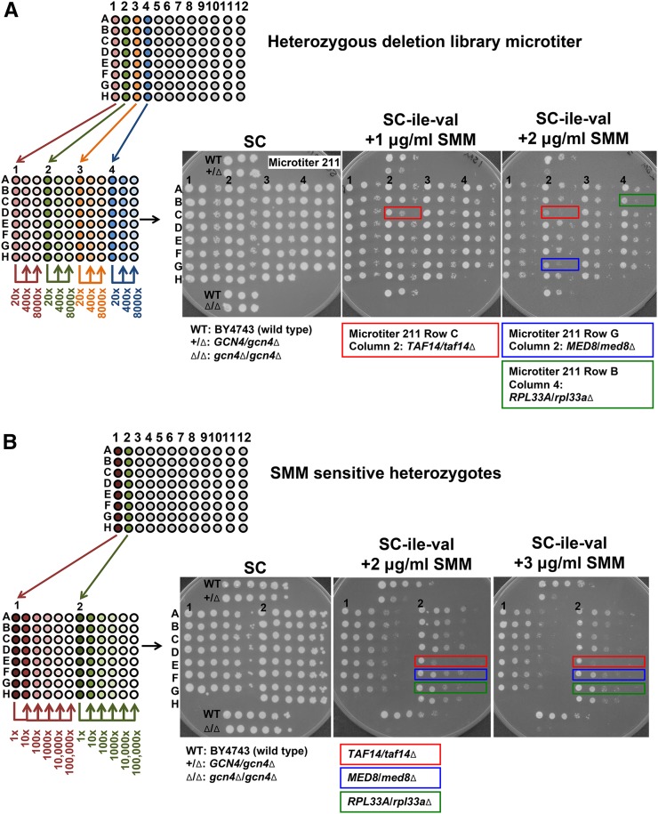
Figure 2.
Screening for strains haploinsufficient for growth in the presence of SMM. Each panel shows a diagram of the dilution series performed as well as plates with representative data. (A) Heterozygous deletion strains from the library microtiters were transferred to microtiters with fresh YPD medium containing G418 sulfate. Every four columns of strains from each library microtiter (numbered) were transferred and diluted 20-fold per new microtiter. Two more 20-fold serial dilutions were made for each strain. For each strain, 5 µl of each dilution (20×, 400×, and 8000×) were spotted onto SC control and SC-ile-val + SMM (1 and 2 µg/ml) agar media. BY4743 (wild type), GCN4/gcn4∆, and gcn4∆/gcn4∆ control strains were grown in separate microtiters in YPD, and diluted samples were included on every agar plate. Plates were photographed after 3, 4, and 5 d of growth. Representative data are shown using the first four columns from microtiter #211 of the heterozygous deletion collection (the photographs show SC and SC-ile-val + 1 µg/ml SMM after 3 d of growth and the SC-ile-val + 2 µg/ml SMM after 4 d of growth). Three strains that displayed significant growth defects in the presence of SMM are indicated: TAF14/taf14∆ (indicated with the red boxes) on both the 1 and 2 µg/ml SMM plates, and MED8/med8∆ (blue box) and RPL33A/rpl33a∆ (green box) on the 2 µg/ml SMM plate. (B) All SMM-sensitive heterozygotes from the library were collected and organized into new microtiters. Two columns from each of the SMM-sensitive candidate microtiters (indicated by numbers 1 and 2 as an example) were used to inoculate YPD + G418 sulfate in fresh microtiters. After 2 d of growth, the strains were serially diluted 10-fold to 100,000× dilution. For each strain, 5 µl of each dilution were spotted onto SC control and SC-ile-val + SMM (1, 2, and 3 µg/ml) agar media. BY4743 (wild type), GCN4/gcn4∆, and gcn4∆/gcn4∆ control strains were grown in separate microtiters in YPD, and diluted samples were included on every agar plate. The photographs shown were taken after 4 d (SC and SC-ile-val + 2 µg/ml SMM) or 5 d (SC-ile-val + 3 µg/ml SMM) of growth. The three strains depicted in (A) are shown here again (the SC-ile-val + 1 µg/ml SMM plate has been omitted for clarity): TAF14/taf14∆ (indicated with the red boxes), MED8/med8∆ (blue boxes), and RPL33A/rpl33a∆ (green boxes).

