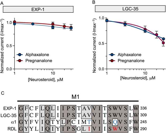
Figure 7.

EXP‐1 and LGC‐35 are not modulated by neurosteroids. (A) EXP‐1 neurosteroid dose–response curves were generated by applying GABA (10 μM) alone or GABA plus increasing concentrations of neurosteroids (10 μM GABA + alphaxalone = 1, 3, 10 and 30 μM; pregnanalone = 1, 3, 10, 20 and 30 μM). Responses were normalized to maximal GABA only responses. Each data point represents the mean ± SEM with n ≥ 6 and N = 2. (B) LGC‐35 neurosteroid dose–response curves were generated by applying GABA (10 μM) alone or with increasing concentrations of neurosteroids (alphaxalone = 1, 3, 10, 30 and 100 μM; pregnanalone = 1, 3, 10, 20 and 30 μM). Responses were normalized to maximal GABA only responses. Each data point represents the mean ± SEM with n ≥ 7 and N = 2. (C) Comparative sequence alignment of transmembrane domain (M1), highlighting key residues that confer differential neurosteroid sensitivity (blue) and resistance (red) in the human α1 GABAA subunit and fly RDL subunit respectively. EXP‐1 and LGC‐35 contain a similar valine and an identical asparagine present in the neurosteroid resistant RDL receptor. Dark grey shading marks amino acid identity and light grey shading identifies similarity.
