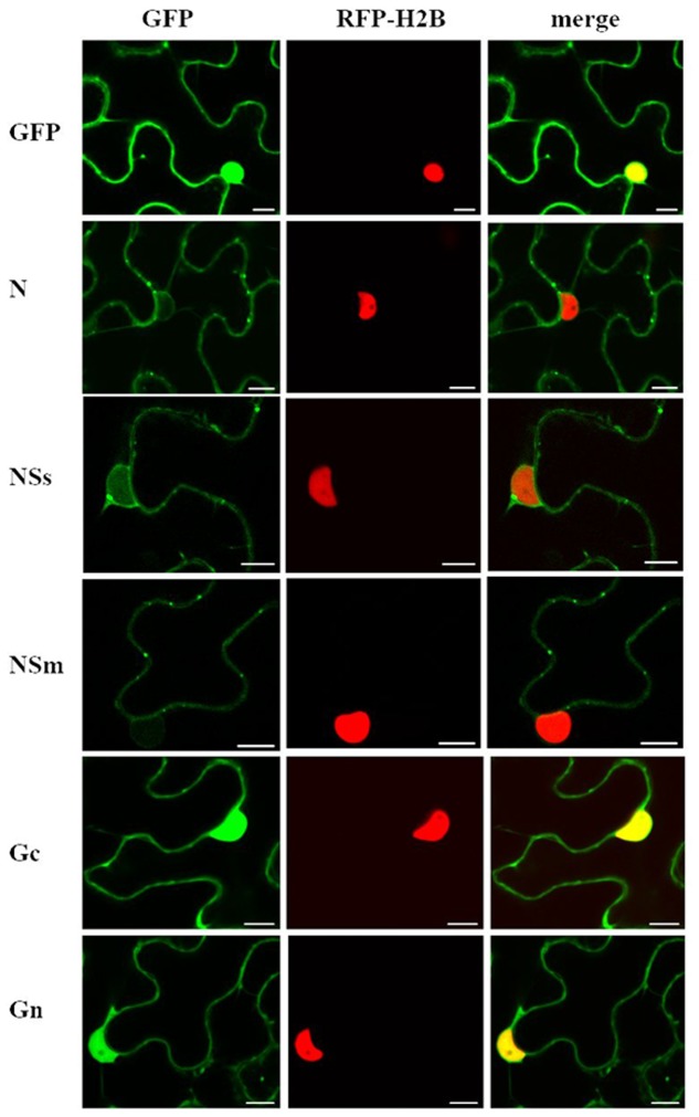
FIGURE 1.
Intracellular localization of transiently expressed free green fluorescent protein (GFP) or capsicum chlorosis virus (CaCV) proteins fused to GFP. CaCV proteins N, NSs, NSm, Gc, Gn were individually expressed from pSITE vectors that were agroinfiltrated into transgenic red fluorescent protein (RFP)-histone 2B (H2B) Nicotiana benthamiana leaf epidermal cells. Images were acquired after 2 days using a confocal microscope at 10 × 25 magnification. Left column, GFP channel; center column, RFP channel; right column, merged images. Bars, 10 μm.

