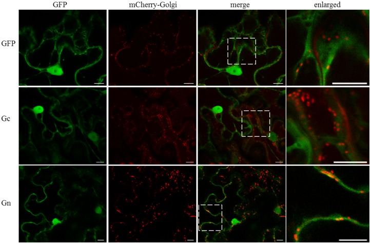
FIGURE 3.
Intracellular localization of free GFP and CaCV Gc and Gn glycoproteins fused to GFP, relative to mCherry-Golgi marker. GFP, viral fusion proteins, and mCherry-Golgi marker were transiently expressed following co-agroinfiltration of gene expression constructs into N. benthamiana leaf epidermal cells. Enlarged sections of images are highlighted with dotted boxes. Images were acquired 2 days after agroinfiltration using a confocal microscope at 10 × 25 magnification. Bar, 10 μm.

