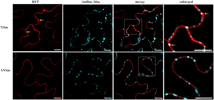
FIGURE 7.
Intracellular localization of CaCV NSm protein and PD. RFP fusions of functional (NSm) and dysfunctional (ΔNSm) protein-expressing N. benthamiana leaves were stained with aniline blue fluorochrome to visualize PD. Enlarged sections of images are highlighted with dotted boxes. Co-localization of NSm aggregates and aniline blue are indicated with arrows. To increase contrast, aniline blue fluorescent images were false colored as cyan. Images were taken at 2 days post infiltration (dpi) using a confocal microscope at 10 × 25 magnification. Bars, 10 μm.

