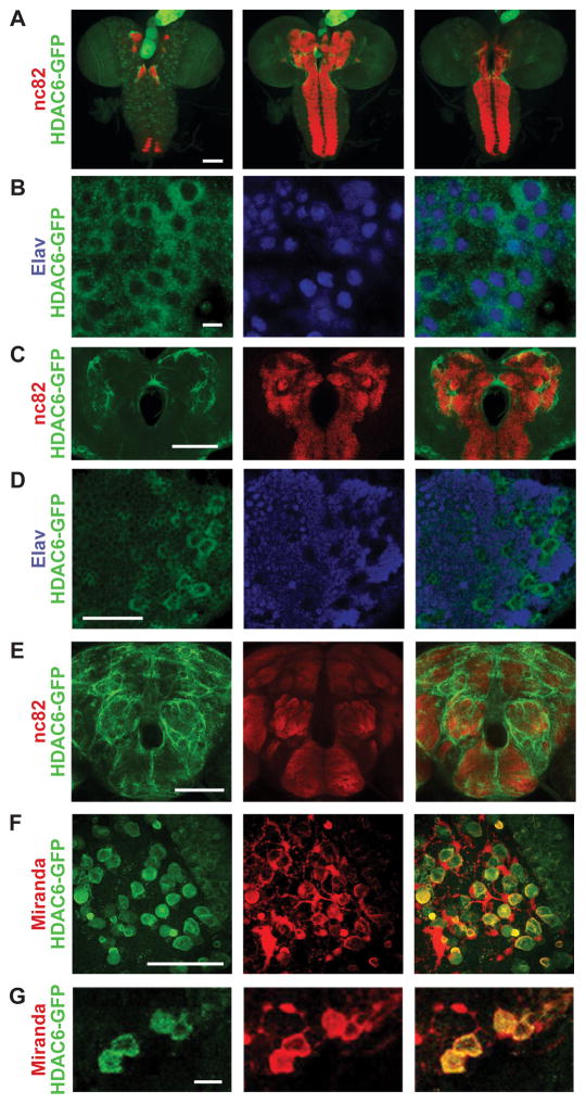
Figure 2. HDAC6 primarily localizes to the cytoplasm in neurons and neuroblasts.
Images showing whole mount antibody staining from an HDAC6-GFP transgenic animal (VK00033{HDAC6-2XTY1-SGFP-V5-preTEV-BLRP-3XFLAG}) where anti-GFP (labeling HDAC6-GFP) is always shown in green.. A. The larval central nervous system (CNS) shown as Z-stacks in three portions going from more dorsal (left), medial (middle) to ventral (right) with anti-Bruchpilot (nc82) is shown in red; B. Larval ventral nerve cord (VNC) with anti-Elav (labeling neuronal nuclei) is shown in blue. White arrows indicate example cell bodies: Neuron with nuclear HDAC6 signal (top); non-neuronal cell with cytoplasmic HDAC6 (middle); Neuron with cytoplasmic HDAC6 (bottom). C. Larval central brain neuropil region with anti-Bruchpilot (nc82) is shown in red. D. Larval optic lobe with anti-Elav (labeling neuronal nuclei) is shown in blue. White arrows indicate putative neuroblasts. E. Adult central nervous system (CNS) with anti-Bruchpilot (nc82) is shown in red; anti-GFP (labeling HDAC6-GFP) is shown in green. F. Larval optic lobe with anti-Miranda (labeling dividing neuroblasts) is shown in red. White arrows indicate putative neuroblasts undergoing asymmetrical cell division with both Miranda and HDAC6 potentially accumulating in the newly forming daughter cell. G. Close-up section of the larval optic lobe showing expression of HDAC6-GFP (green) in anti-Miranda labeled neuroblasts (red). Scale bars correspond to 100μm: (A, E); 50μm (C, D, F); 10μm (B, G).

