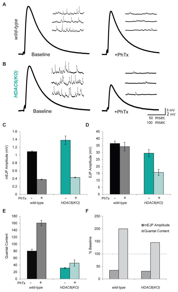
Figure 4. Homeostatic synaptic plasticity is disrupted in HDAC6 mutants.
A. Representative electrophysiological traces showing evoked excitatory junctional potentials (EJP) and mini excitatory junctional potentials (mEJPs) from wild-type larva before and after PhTx treatment. B. Representative traces from HDAC6(KO) larva before and after PhTx treatment. C. Average mEJP amplitudes in wild-type and HDAC6(KO) larva with and without PhTx treatment. D. Average EJP amplitudes in wild-type and HDAC6(KO) larva with and without PhTx treatment. E. Estimated quantal content in wild-type and HDAC6(KO) larva with and without PhTx treatment. F. mEJP amplitude and quantal content in wild-type and HDAC6(KO) larva following PhTx treatment shown as percent base line. (N = 10–14) G. Model for HDAC6’s role in synaptic plasticity: in wild-type animals, HDAC6 may deacetylate BRP C- terminal domains to increase vesicle tethering during synaptic strengthening. This would result in increased quantal content and increased transmission at these synapses. In HDAC6 mutant animals, BRP would not be deacetylated during synaptic strengthening. This may result in a limited increase in quantal content and a deficit in plasticity. Error bars represent +- s.e.m

