
FIGURE 2.
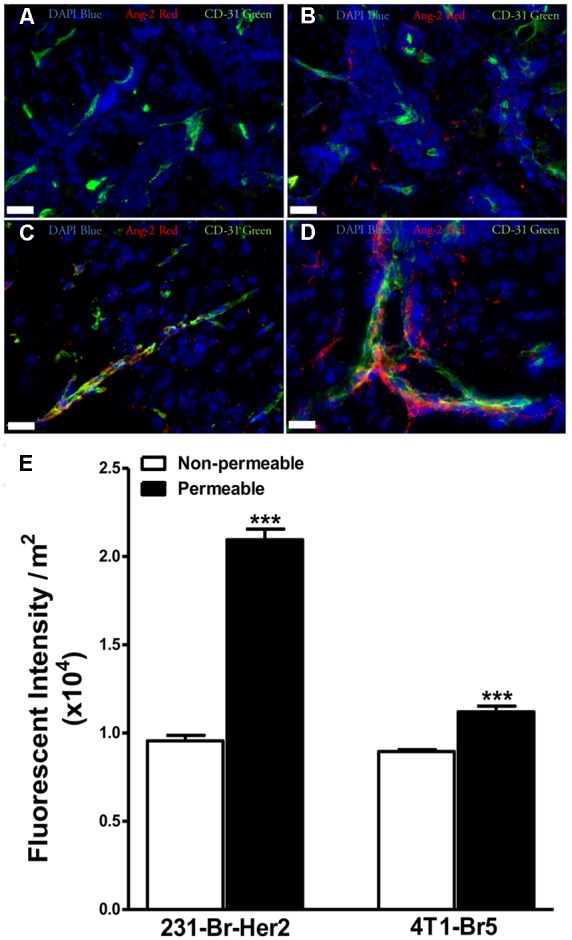
Expression of angiopoietin-2 is greater in lesions with higher permeability. Brain slices from un-treated metastases bearing animals were stained for the hypoxia induced vascular destabilizing protein Ang-2. Ang-2 expression in MDA-MB-231-BR-Her2 metastases is shown in non-permeable (A) and permeable (B) metastatic lesions. Representative images in the 4T1-BR5 model are shown in a lesion with low permeability (C) and a lesion with high permeability (D). Ang-2 (red), CD31 (green), DAPI (blue). Ang-2 intensity in permeable and non-permeable lesion in both preclinical models of brain metastases of breast cancer (E). Permeable lesions demonstrated greater Ang-2 expression than non-permeable in both models, with a >2-fold increase in the MDA-MB-231-BR-Her2 model, indicating the presence of vascular destabilization. Scale bar = 25 μm. Statistical significance was determined using Student’s t-test to determine difference in Ang-2 expression between the two permeability groups within each model (∗∗∗ for p < 0.001).