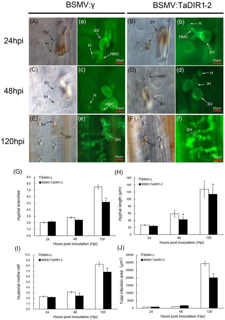
Figure 7.
Histological observation of fungal growth and necrotic cell area in wheat infected with BSMV:γ or BSMV:TaDIR1-2 after inoculation with the Pst virulent pathotype CYR31. The fungal growth and necrotic cell area in wheat leaves inoculated with BSMV:γ or BSMV:TaDIR1-2at 24 hpi (Aa,Bb), at 48 hpi (Cc,Dd), and at120 hpi (Ee,Ff) were observed under a light microscope. The same letter indicates the photo was taken at the same infection site. The average number of hyphal branch and haustorial mother cell (G,I) of fungus in each infection site was counted. (H) Hyphal length, (the average distance from the junction of the substomatal vesicle and the hypha to the tip of the hypha), was measured using DP-BSW software (unit in μm). (J) Infection area (the average area of the expanding hypha) was calculated using DP-BSW software (units of 103 μm2). All results were obtained from 50 infection sites. Asterisks indicate a significant difference (P < 0.05) from the BSMV:γ by Student's t-test. HMC, haustorial mother cell; HR, hypersensitive reaction; IH, infection hypha; SH, secondary hypha; SV, substomatal vesicle; HMC, hostorial mother cell; H, haustorium.

