Figure 4.

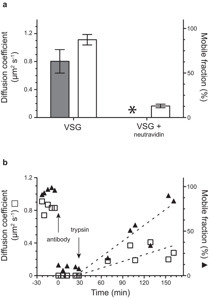
Cross-linking of VSG in supported membranes. a) As a control for the mSAV experiment, tetravalent neutravidin was added to biotinylated VSG. This leads to rapid crosslinking and immobilization of the protein complexes. The mobile fraction  was reduced 7-fold and no diffusion coefficient
  could be calculated using Soumpasis diffusion algorithm for complexed proteins
  . The diffusion coefficient
  and mobile fraction
  of untreated VSG was normal. Data are means ± SD. b) Treatment of VSG in supported membranes with a VSG-specific antibody causes immediate immobilization of the protein complexes that is slowly reversed after trypsin digestion. Arrows mark the addition of rabbit anti-VSG M1.6 antibody and trypsin, respectively. Diffusion coefficient (□), mobile fraction
  . The dotted lines represent linear regression and are guidelines for the eye.
