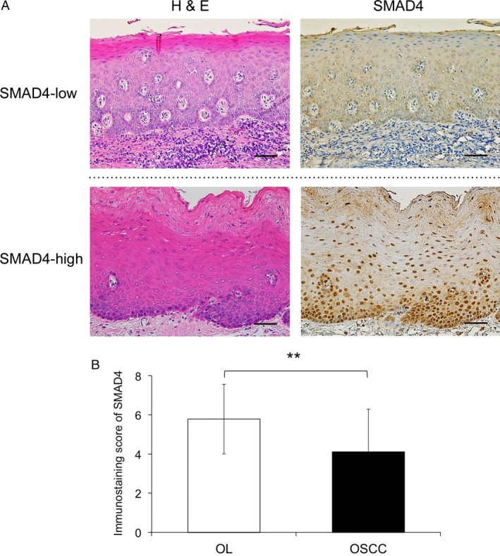
Figure 1.

Immunohistochemical staining of SMAD4 in oral leukoplakia and oral squamous cell carcinoma tissues. (A) Representative immunohistochemical staining patterns of SMAD4 are shown according to the expression status. The sections were stained with hematoxylin and eosin (H&E) and anti‐SMAD4 polyclonal antibodies. In the oral leukoplakia (OL) specimens, SMAD4 was mainly expressed in the nucleus of the epithelial cells. Original magnification, ×200, scale bar = 100 μm. (B) Immunostaining scores obtained from each pathological group were calculated and statistically analyzed by the Mann–Whitney U test. The y axis shows the mean values of the SMAD4 immunostaining scores. **, P < 0.01 oral squamous cell carcinoma, OSCC.
