
Figure 2.
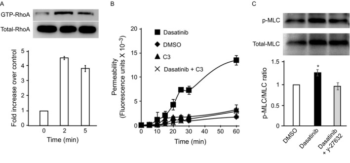
RhoA activation in dasatinib‐treated cells. (A) Rhotekin‐linked agarose beads were used to pull down Rho‐GTP from lysates of human microvascular endothelial cells‐1 (HMEC‐1) at indicated times after dasatinib incubation. The samples were then immunoblotted with anti‐RhoA Ab. (B) Effect of RhoA inhibition on endothelial permeability. HMEC‐1 were seeded onto Transwell insert and incubated with vehicle [dimethyl sulfoxide (DMSO)], dasatinib or the combination of dasatinib and the RhoA inhibitor I, C3. At various times, the lower chamber was sampled and analyzed for the presence of fluorescein isothiocyante‐dextran. Results are mean and SD of quadruplicate wells of typical experiments. (C) HMEC‐1 were incubated with dasatinib (100 ng/mL) and the cells were solubilized. Lysates were subjected to SDS‐PAGE, electrophoretically transferred to PVDF membranes and immunoblotted with antibodies to total and Phospho‐myosin light chain. The ratios of relative band intensities (means and SD) of three independent experiments are shown. * denotes a P value of less than 0.05 compared to DMSO.