
Figure 3.
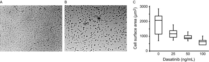
Effect of dasatinib on endothelial cell morphology. Human microvascular endothelial cells‐1 were grown on fibronectin‐coated coverslips. Following confluence (24–48 h), the cells were incubated with dimethyl sulfoxide (A) or dasatinib (B) for 60 min, and examined under microscope (100×). In (C), cell suspensions were incubated with various concentrations of dasatinib (60 min), allowed to adhere to fibronectin‐coated plates for 30 min at 37°C. The cells were fixed, stained with eosin, and cell surface area was quantified by ImageJ software.