
Figure 6.
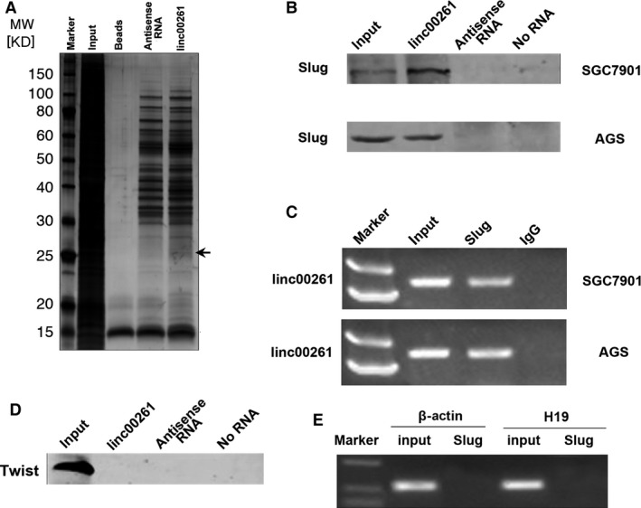
linc00261 binds to Slug protein. (A) Representative SDS‐PAGE gel after Coomassie blue staining shows a prominent band at about 25 kD, representing mainly Slug proteins (see also supplemental Table 4). (B) RNA pull‐down assay performed in SGC7901 and AGS cells. Biotinylated linc00261 or antisense RNA was incubated with cell extracts, targeted with streptavidin beads and washed, and the associated proteins were resolved on a gel. Western blot analysis detected the specific association of Slug and linc00261 (n = 3). (C) RIP experiments were performed using the Slug antibody for immunoprecipitation (IP) and a primer to detect linc00261. RIP enrichment was determined relative to the input controls. (D) No interaction between linc00261 and Twist could be detected. RNA pull‐down assay performed in SGC7901 cells. Biotinylated linc00261 or antisense RNA was incubated with cell extracts, targeted with streptavidin beads and washed, and the associated proteins were resolved on a gel. Western blot analysis detected the specific association of Twist and linc00261. (E) RIP experiments were performed using Slug to immunoprecipitate RNA and a primer to detect H19 and β‐actin.