
Figure 7.
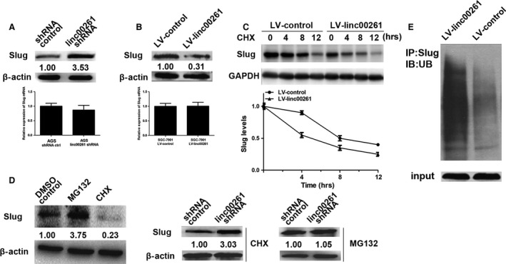
linc00261 decreases the protein level of Slug by inhibiting its stability. (A) The protein levels of Slug were detected in linc00261‐knockdown AGS cells by Western blot analysis. The mRNA levels of Slug were detected in linc00261‐knockdown AGS cells by qRT‐PCR (n = 3). (B) The protein levels of Slug were detected in linc00261‐up‐regulated SGC7901 cells. The mRNA levels of Slug were detected in linc00261‐overexpressing SGC‐7901 cells by qRT‐PCR (n = 3). (C) linc00261 overexpression enhanced Slug protein degradation. SGC‐7901 cells were transfected with Luc or linc00261. After treating the cells with cycloheximide (CHX, 0.5 μg/μl) for an indicated time, the expression of endogenous Slug protein was analysed by WB. The band intensity of Slug for each time‐point was quantified by ImageJ and plotted. Experiments were repeated for three times, and a representative experiment is presented. Error bars represent S.D. Every experimental group was compared with the control Lucsi group. (D) The comparison and quantification of Slug proteins in AGS cells with and without the protein synthesis inhibitor cycloheximide (CHX, 0.5 μg/μl) or the proteasome inhibitor MG‐132 (5 μM) for 24 hrs. linc00261 stable knockdown AGS cells and control cells were incubated with MG132 (5 μM) or CHX (0.5 μg/μl) for 24 hrs. The levels of Slug proteins were detected by Western blot analysis. (E) Stable linc00261 overexpressing SGC7901 cells and control cells were treated with MG132 (10 mM) for 6 hrs. Cell lysates were immunoprecipitated with a Slug‐specific antibody, followed by Western blotting with an antibody to ubiquitin. The bottom panel depicts the input of the cell lysates.