Figure 2.

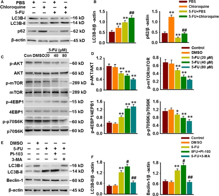
5‐FU induces autophagosome formation through PI3K/AKT/mTOR pathway. (A) HepG2 cells were pre‐treated with PBS or chloroquine (1 μM) for 30 min. and were then exposed 5‐FU (80 μM) for 48 hrs. LC3B and p62 expressions were detected by Western blotting. (B) Densitometric analysis of LC3B‐II and p62 protein expression. **P < 0.01 versus PBS, ##P < 0.01 versus 5‐FU+PBS, n = 5. (C) Western blotting images showing the phosphorylation of AKT, mTOR, 4EBP1 and p70S6K in HepG2 cells after 5‐FU treatment for 48 hrs. (D) Densitometric analysis of these protein expression was performed. *P < 0.05, **P < 0.01 versus DMSO, n = 6. (E) Cells were pre‐treated with PI‐103 (0.5 μM) or 3‐MA (5 μM) prior to incubation with 5‐FU (80 μM) for 48 hrs. Western blotting analysis of LC‐3B and Beclin‐1 expression. (F) Bar diagram represents the densitometric analysis of LC3B‐II and Beclin‐1. **P < 0.01 versus DMSO, #P < 0.05, ##P < 0.01 versus 5‐FU, n = 4.
