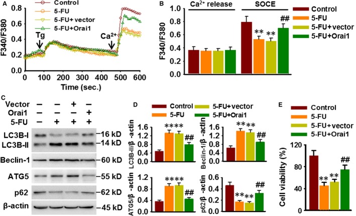
Figure 6.

Restoration of Orai1 attenuates 5‐FU‐induced autophagic cell death. (A) Cells were transfected with Orai1 plasmid for 48 hrs prior to 5‐FU (80 μM) incubation for another 48 hrs. Ca2+ imaging experiment was performed as mentioned in methods section. (B) Quantification of fluorescence ratio (340/380). **P < 0.01 versus control, ##P < 0.01 versus 5‐FU, n = 6. (C) Representative Western blotting images showing the expression of LC3B, Beclin‐1, ATG5 and p62. (D) Densitometric analysis was performed using ImageJ analysis software. **P < 0.01 versus control, ##P < 0.01 versus 5‐FU, n = 5. (E) Cell viability was examined by CCK‐8 assay. **P < 0.01 versus control, ##P < 0.01 versus 5‐FU, n = 6.
