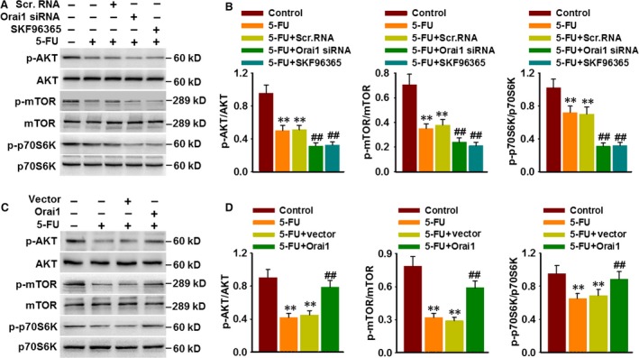
Figure 7.

Orai1 inhibits 5‐FU‐induced autophagy through AKT/mTOR signalling pathway. (A) HepG2 cells were treated with Orai1 siRNA (50 nM) for 48 hrs or treated with SKF96365 (20 μM) for 3 hrs prior to 5‐FU (80 μM) incubation for another 48 hrs. The phosphorylation of AKT, mTOR and p70S6K were determined by Western blotting. (B) Bar diagram represents the densitometric analysis of the phosphorylation level of above proteins. **P < 0.01 versus control, ##P < 0.01 versus 5‐FU, n = 5. (C) Cells were transfected with Orai1 plasmid for 48 hrs and then were treated with 5‐FU (80 μM) for another 48 hrs. Representative Western blotting images showing the phosphorylation of AKT, mTOR and p70S6K. (D) Densitometric analysis of AKT, mTOR and p70S6K phosphorylation was performed. **P < 0.01 versus control, ##P < 0.01 versus 5‐FU, n = 6.
