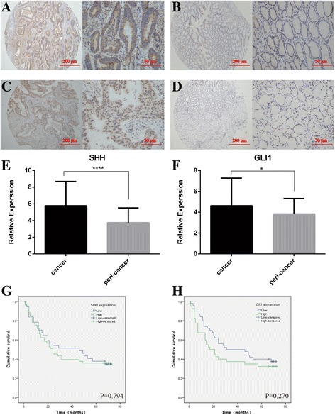
Fig. 4.

Immunostaining analysis of the Hh signaling molecules (SHH and GLI1) in human gastric cancer tissues. SHH expression is mainly expressed in the cytoplasm and GLI1 protein is strongly localized to the nucleus and cytoplasm in gastric cancer cells. The micrographs showed representative immunohistochemical staining of SHH (a) and GLI1 (c) in the gastric cancer tissues. The expression of SHH and GLI1 in corresponding adjacent normal gastric tissues is shown in (b) and (d) respectively (magnification: left panel × 100, right panel × 400). Quantitative analysis of SHH (e) and GLI1 (f) in tumor tissues and adjacent normal gastric tissues. *p < 0.05 and ***P <0.001 for SHH and GLI1 vs their respective controls, respectively. The differences in overall survival time between high and low expression of SHH (g) and GLI1 (h) in gastric cancer tissue are determined using the Kaplan-Meier method
