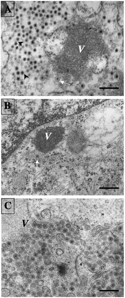
FIG. 5.
Morphogenesis of RRV rotavirus particles in MA104 cells transfected with siRNAs. MA104 cells transfected with (A) the siRNA control (siRNActrol), (B) siRNANSP4, or (C) siRNAVP7 were infected 48 h posttransfection, and at 8 hpi, the cells were fixed and prepared for electron microscopy as indicated in Materials and Methods. Dense viroplasmic inclusions (V) are present in the cytoplasm of rotavirus-infected cells, adjacent to the ER. From these structures, DLPs bud into the lumen of the ER, resulting in membrane-enveloped particles (arrows) which later lose the membrane to produce mature triple-layered virions (arrowheads). The pictures shown are representative of at least 20 different virus-infected cells. Magnification, ×15,000. Scale bars, 400 nm.

