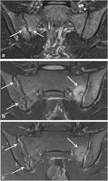
Fig. 2.

Active sacroiliitis in a 16-year-old girl with juvenile spondyloarthritis according to global assessment as well as to the ASAS definition of a positive MRI for sacroiliitis. a Semicoronal STIR image shows two small, focal spots of BME at the sacral and iliac side of the right sacroiliac joint (arrows). b Follow-up MRI 6 months later shows more extensive active sacroiliitis with bilateral high signal in the joint space and an active lesion with surrounding BME at the sacral side of the left sacroiliac joint (arrows). c Corresponding semicoronal fat-saturated T1-weighted image of the follow-up MRI shows bilateral enhancement of the synovium (synovitis) and of the active lesion on the left side (arrows)
