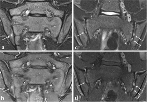
Fig. 3.

Active sacroiliitis in a 14-year-old (left) and 13-year-old (right) boy with juvenile spondyloarthritis according to a global assessment of MRI for sacroiliitis, not according to the ASAS definition of a positive MRI for sacroiliitis. Semicoronal STIR (a and c) and contrast-enhanced fat-saturated T1-weighted (b and d) images showing on the left side a focal enhancing BME lesion (seen on only one slice) at the iliac side of the right sacroiliac joint (arrows), and on the right side showing bilateral multiple enhancing spots of nodular high signal in the joint space, representing active erosions (arrows). No BME is seen. Note also the enlarged para-iliacal lymph nodes (asterisks)
