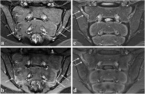
Fig. 4.

Active sacroiliitis in a 14-year-old girl (left) and a 14-year-old boy (right) with spondyloarthritis according to a global assessment of MRI for sacroiliitis, not according to the ASAS definition of a positive MRI for sacroiliitis. Semicoronal STIR (a and c) and contrast-enhanced fat-saturated T1-weighted (b and d) images on the left side showing synovitis in the caudal part of the left SI joint, also discrete in the caudal part of the right SI joint, seen as high signal in the joint space on STIR with corresponding synovial enhancement. Bone marrow edema is absent. On the right side, synovitis/retro-articular enthesitis is shown at the right sacroiliac joint (arrows). No BME is seen
