
Fig. 3.
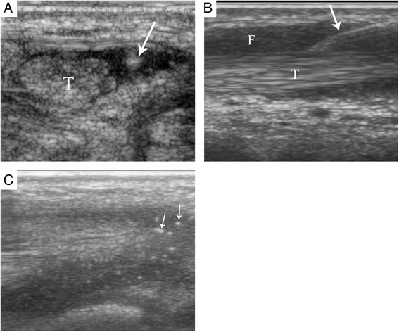
Sonographic images of tendon sheath injection approach. a An out of plane injection of the tibialis posterior tendon sheath of a 13 years 8 months old male. b An in plane injection of the tibialis posterior tendon sheath of a 9 years 11 months old female. Arrows point to the needle. c Steroid solution (small arrows) surrounding the tibialis posterior tendon of a 9 year 4 months old male after injection. T = Tendon, F = Peritendinous fluid