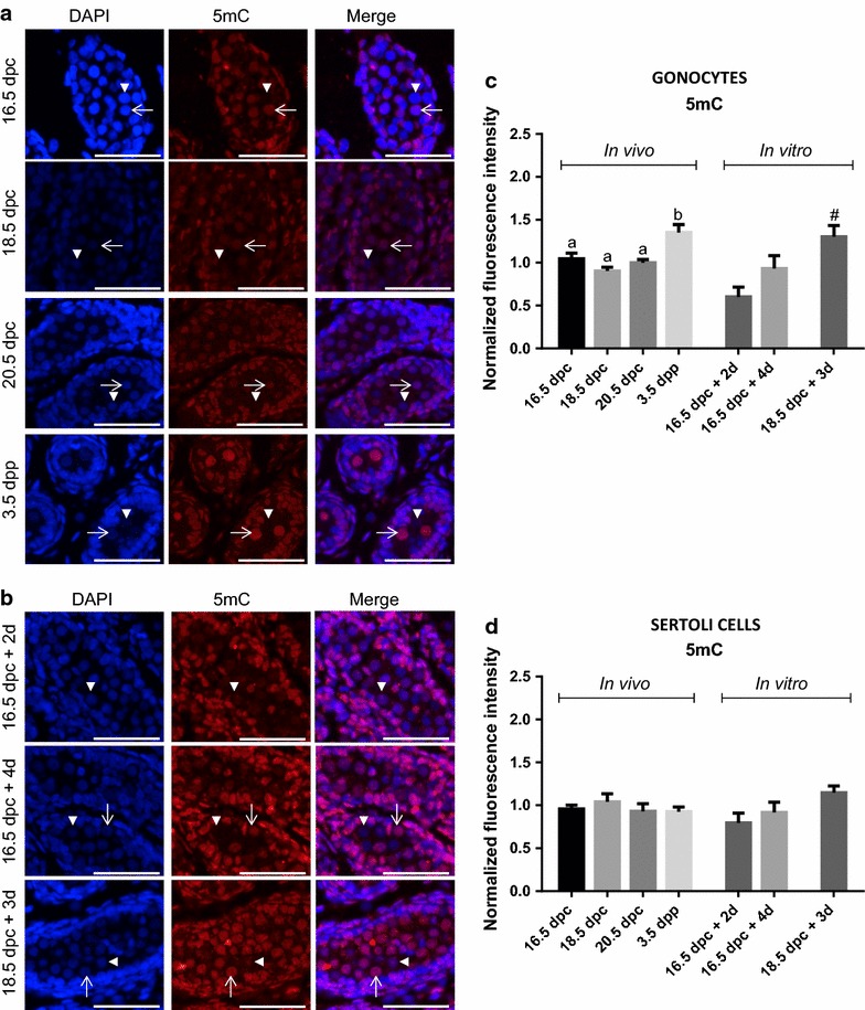
Fig. 4.

Global changes in DNA methylation in gonocytes and Sertoli cells in vivo and in vitro. Co-staining of 5mC (red) and DAPI (blue) was done on sections of rat testes sampled in vivo (a) or after organ culture (b). Note that at all age, we observed both unmethylated (arrow head) and methylated gonocytes (arrow). Immunofluorescence intensity was quantified in gonocytes (c) and in Sertoli cells (d). Data represent the average normalized fluorescence intensity ± SEM (n = 3/time point). a,b p < 0.05 between different stages in vivo using a one-way ANOVA followed by a Tukey’s post hoc test. # p < 0.05 between 18.5 dpc and after culture using a Student unpaired t test. Scale = 50 μm
