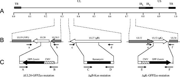
FIG. 1.
Schematic of the strategy for the construction of pYEbac102 mutant BACs. (A) The top line represents the prototypic arrangement of the HSV-1 genome, with the unique long (UL) and unique short (US) regions flanked by the terminal repeat (TR) and internal repeat (IR) regions. (B) Shown below are the expanded genomic regions of the UL20, UL27, and UL53 ORFs as well as the approximate locations of the sites to which insertion of the marker genes was targeted and the primers used in diagnostic PCR to confirm the presence of each mutation. (C) PCR fragments containing the kanamycin resistance or GFP-zeocin resistance gene cassette flanked by ≈50 bp of viral sequences on both sides were used for targeted GET recombination in E. coli to construct pYEbac102 mutant BACs with insertion-deletion mutations in the UL20, UL27, and/or UL53 ORFs. The approximate locations of the primers used in amplification of each PCR fragment are also shown.

