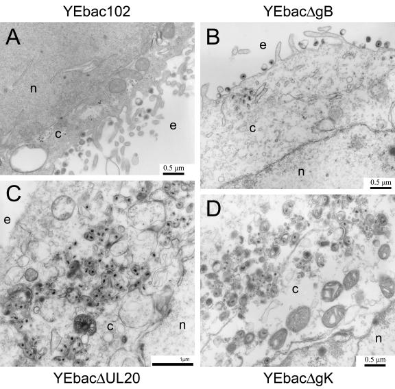
FIG. 6.
Ultrastructural morphology of gB-, gK-, and UL20-null viruses. Electron micrographs of Vero cells infected with YEbac102 (A), YEbacΔgB (B), YEbacΔUL20 (C), or YEbacΔgK (D). Confluent cell monolayers were infected at an MOI of 5, incubated at 37°C for 24 h, and prepared for transmission electron microscopy. Nuclear (n), cytoplasmic (c), and extracellular (e) spaces are marked.

