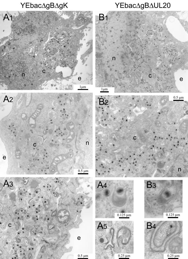
FIG.7.
Ultrastructural morphology of gB/gK and gB/UL20 double-null viruses. Electron micrographs of Vero cells infected with YEbacΔgBΔgUL20 (A1, A2, A3, A4, and A5) or YEbacΔgBΔgK (B1, B2, B3, and B4). Confluent cell monolayers were infected at an MOI of 5, incubated at 37°C for 24 h, and prepared for transmission electron microscopy. Panels A4 and B3 show partially enveloped capsids. Panels A5 and B4 show a tegument-like accumulation on membranes that are folded irregularly. Nuclear (n), cytoplasmic (c), and extracellular (e) spaces are marked.

