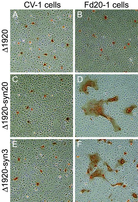
FIG. 9.
Plaque phenotypes of Δ1920, Δ1920-syn20, and Δ1920-syn3 viruses on CV-1 (A, C, and E) and FcgK-1 (B, D, and F) cells. Confluent cell monolayers were infected with the Δ1920 (A and B), Δ1920-syn20 (C and D), or Δ1920-syn3 (E and F) at an MOI of 0.001, and viral plaques were visualized at 48 h postinfection by immunohistochemistry.

