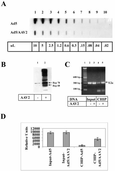
FIG. 3.
Rep proteins are cross-linked to the Ad5 E2a promoter region during AAV2 coinfection. (A) Equal amounts of DNA from formaldehyde-cross-linked nuclei from Ad5-infected or Ad5- and AAV-coinfected HeLa cells were analyzed by slot blot hybridization with an Ad5 probe. The numbers at the bottom of the panel refer to the amount of purified DNA from the original 50 μl obtained after chromatin isolation. (B) The presence of the Rep proteins was verified by SDS-PAGE and immunoblot analysis after CHIP analyses. Lane 1 is from Ad-infected cells, and lane 2 is from Ad-AAV-coinfected cells. (C) CHIP analysis was performed with primers that amplify the Ad5 E2a region. Equivalent amounts of input (lanes 2 and 3) and CHIP (lanes 4 and 5) Ad DNA (as determined in panel A) were amplified and separated by agarose gel electrophoresis. Lane 1 contains 1.0 μg of 100-bp DNA ladder separated. (D) The ethidium-bromide stained bands from panel C were quantitated by using a Kodak Image Station 440 from PCRs performed in triplicate. Error bars represent standard deviations.

