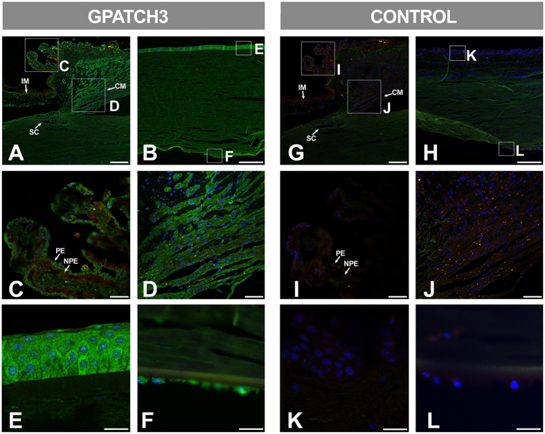
Figure 3. Fluorescent immunohistological detection of GPATCH3 in anterior segment tissues of a human eye.
(A–L) Fluorescent immunohistochemistry of histological sections (10 μm) of a human eye from a 45-year-old Caucasian female donor (cadaver) with no reported ocular pathology. Samples were incubated with either rabbit anti-GPATCH3 primary antibody and Cy2 donkey anti-rabbit secondary antibody (A–F) or only secondary antibody as a negative control (G–L). Confocal wide-field micrographs of iridocorneal angle (A and G) or cornea (B and H) and detailed images of ciliary processes (C and I), ciliary muscle (D and J), corneal epithelium (E and K) and endothelium (F and L) are shown. Scale bars indicate 200 μm in panels A, B, G and H, 50 μm in panels C, G, I and J and 20 μm in panels E, F, K and L. The green channel corresponds to GPATCH3, the red channel to tissue autofluorescence and the blue channel to DAPI nuclear staining. IM: Iris muscle. CM: Ciliary muscle. SC: Schlemm channel. PE: Pigmented epithelium. NPE: Non-pigmented epithelium.

