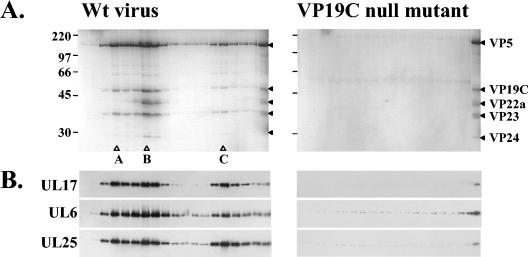
FIG. 2.
Sucrose gradient sedimentation analysis of extracts from BHK cells infected with wt virus strain 17 or the VP19C null mutant vΔ38YFP. The extracts were sedimented through a 10 to 40% sucrose gradient, and 32 successive fractions were collected starting from the top of the gradient. The proteins in fractions 14 to 32 were analyzed by SDS-PAGE and detected by staining with Coomassie brilliant blue (A). Protein size markers (in kilodaltons), indicated by the short lines, are shown at the far left-hand side of the panel. The open triangles show the peak fractions of A, B, and C capsids. The far lane on the right-hand side of each image of the gel shows a wt B-capsid protein profile. The HSV-1 capsid proteins are denoted by the closed triangles. The packaging proteins in fractions 14 to 31 were identified by Western blotting, using UL6, UL17, and UL25 antibodies (B).

