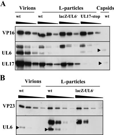
FIG. 8.
Comparison of UL6 in wt virions and wt and mutant L particles. Serial twofold dilutions of virions and L particles were prepared, and the proteins were separated by SDS-PAGE. Proteins were detected by Western blotting using antibodies against UL6, UL17, VP16, and VP23. Virions and L particles were equalized on the basis of their VP16 content (A). A sample of wt B capsids was also included to distinguish UL6 from cross-reacting proteins. For panel B, wt virions and L particles were equalized on the basis of their VP23 content to determine whether capsid contamination of L particles accounted for the presence of UL6. UL6 protein is indicated by a closed triangle.

