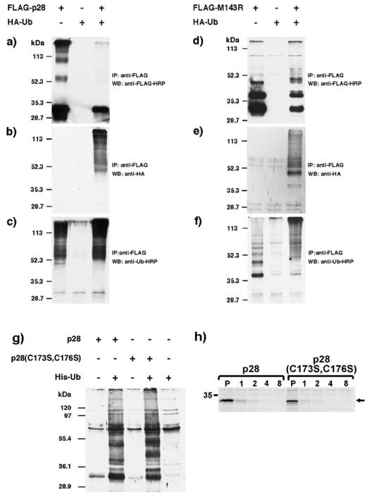
FIG. 3.
Ubiquitination of p28 and M143R. CV-1 cells were infected with the indicated recombinant viruses at an MOI of 5. Sixteen hours postinfection, cells were treated with 25 μM MG132 for 4 h, lysed in RIPA buffer, and immunoprecipitated with anti-FLAG antibody (M2; Sigma). Immunoprecipitates were separated by sodium dodecyl sulfate-polyacrylamide gel electrophoresis (SDS-PAGE) and were blotted with either anti-FLAG-horseradish peroxidase (a and d), anti-HA (b and e), or anti-ubiquitin-horseradish peroxidase (c and f). (g) CV-1 cells were infected with the indicated viruses at an MOI of 2 for 16 h, treated with 25 μM MG132 for 4 h, and lysed in 1% NP-40. Cleared lysates were incubated with Ni2+ resin for 2 h at 4°C, and bound proteins were eluted using 1 M imidazole in 500 mM NaCl and 20 mM Tris (pH 7.9) and concentrated by acetone precipitation. Samples were separated by SDS-PAGE and probed with anti-FLAG-HRP (M2). (h) HeLa cells were infected with the indicated viruses at an MOI of 5 for 16 h and were metabolically labeled with [35S]methionine (0.1 mCi/ml) for 30 min, and the label was chased for the indicated hours. Cells were lysed, and p28 was immunoprecipitated using anti-FLAG. WB, Western blot; IP, immunoprecipitation; Ub, ubiquitin; HRP, horseradish peroxidase.

