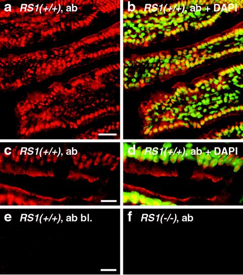
FIG. 2.
Distribution of RS1 protein in the small intestines of C57BL/6J wild-type mice. Cryosections of the jejunum from 5-month-old RS1+/+ mice (a to e) and RS1−/− mice (f) were fixed with paraformaldehyde and incubated with affinity purified RS1 antibody (ab). The immunoreaction was visualized with Cy3-coupled secondary antibody against rabbit IgG F(ab′)2 (red fluorescence). Nuclei were counterstained with DAPI (b and d) (blue fluorescence that was converted to green). In panel e, the immunoreaction was blocked with the antigenic peptide (bl.). In panels b and d, the nuclear localization of RS1 is demonstrated by colocalization of red Cy3 fluorescence of RS1 antibody and green DAPI staining of the nuclei. Colocalizations show up in yellow. Bars, 20 μm (a); 5 μm (c and e).

